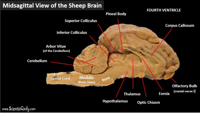
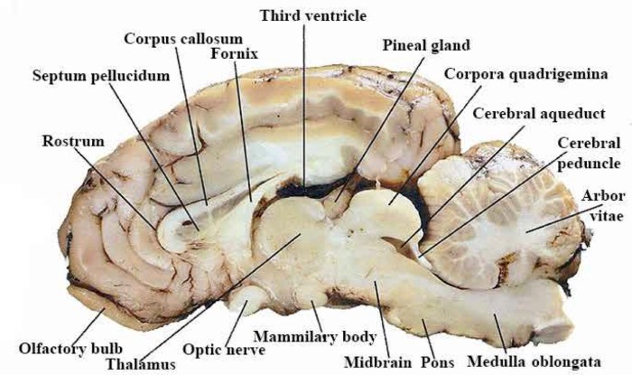
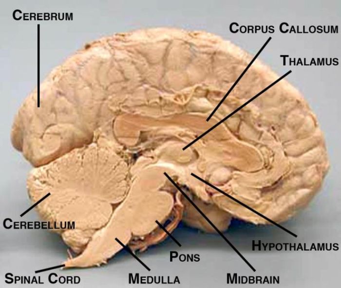
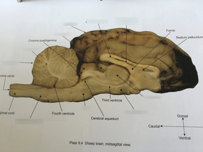
The sheep brain midsagittal view labeled provides a detailed examination of the brain’s internal structures, offering valuable insights into its anatomy and function. This intricate diagram allows for the identification of key brain regions and their corresponding roles, facilitating a deeper understanding of the brain’s complex mechanisms.
The midsagittal view dissects the brain along its midline, revealing the intricate arrangement of brain structures within the cranial cavity. By examining this view, researchers and medical professionals gain a comprehensive perspective on the brain’s organization and the relationships between its various components.
General Overview
The midsagittal view of the sheep brain is a section of the brain that has been cut in the median sagittal plane, dividing the brain into left and right halves. This view provides a comprehensive look at the internal structures of the brain, including the cerebrum, cerebellum, brainstem, and ventricles.
Labeled Diagram
[Image of a sheep brain in the midsagittal view with labels]
Major Structures

The midsagittal view of the sheep brain reveals several major structures involved in various neurological functions. These structures include the cerebral hemispheres, diencephalon, midbrain, pons, medulla oblongata, and cerebellum.
Major Structures and Functions
| Structure | Function | Brain Region |
|---|---|---|
| Cerebral Hemispheres | Higher-level cognitive functions, sensory processing, motor control | Forebrain |
| Diencephalon | Relay sensory information, regulate body functions | Forebrain |
| Midbrain | Relay sensory and motor information, control eye movements | Midbrain |
| Pons | Relay sensory and motor information, control respiration | Hindbrain |
| Medulla Oblongata | Control vital functions (breathing, heart rate), relay sensory and motor information | Hindbrain |
| Cerebellum | Coordination of movement, balance, and posture | Hindbrain |
White Matter Tracts
The white matter tracts are bundles of myelinated axons that connect different regions of the brain. They are named according to their origin and destination. In the midsagittal view of the sheep brain, several major white matter tracts are visible.
Major White Matter Tracts
- Corpus callosum:The corpus callosum is a large white matter tract that connects the two cerebral hemispheres. It allows for communication between the two hemispheres and plays a role in higher-order cognitive functions such as language and memory.
- Internal capsule:The internal capsule is a white matter tract that lies between the thalamus and the basal ganglia. It contains fibers that connect the cerebral cortex to the brainstem and spinal cord.
- Cerebral peduncles:The cerebral peduncles are two white matter tracts that connect the cerebrum to the midbrain. They contain fibers that carry motor and sensory information between the cerebrum and the rest of the body.
- Medial lemniscus:The medial lemniscus is a white matter tract that carries sensory information from the body to the thalamus. It is located in the brainstem.
- Spinothalamic tract:The spinothalamic tract is a white matter tract that carries pain and temperature information from the body to the thalamus. It is located in the brainstem.
Ventricular System

The ventricular system consists of a series of interconnected cavities within the brain that are filled with cerebrospinal fluid (CSF). In the midsagittal view of the sheep brain, the following ventricles are visible:
- Lateral ventricles: These are the largest ventricles and are located within the cerebral hemispheres. They are connected to the third ventricle via the interventricular foramina.
- Third ventricle: This is a narrow, slit-like ventricle located between the thalamus and hypothalamus. It is connected to the lateral ventricles via the interventricular foramina and to the fourth ventricle via the cerebral aqueduct.
- Fourth ventricle: This is a diamond-shaped ventricle located at the base of the brainstem. It is connected to the third ventricle via the cerebral aqueduct and to the subarachnoid space via the median and lateral apertures.
The ventricular system plays a vital role in the production, circulation, and absorption of CSF. CSF is a clear, colorless fluid that bathes the brain and spinal cord and provides them with nutrients and oxygen.
Cerebellum and Brainstem
In the midsagittal view of the sheep brain, the cerebellum and brainstem are clearly visible. The cerebellum, located posterior to the cerebrum, plays a crucial role in coordinating movement and maintaining balance. The brainstem, connecting the cerebrum to the spinal cord, consists of the midbrain, pons, and medulla oblongata.
These structures are responsible for a wide range of vital functions, including respiration, heart rate, and consciousness.
Cerebellum
- Coordinates movement and maintains balance
- Receives sensory input from the body and sends motor commands to the muscles
- Contributes to cognitive functions such as attention, planning, and language
Brainstem
- Consists of the midbrain, pons, and medulla oblongata
- Controls vital functions such as respiration, heart rate, and consciousness
- Relays sensory and motor information between the cerebrum and the spinal cord
Functions of the Cerebellum and Brainstem:
- Motor coordination and balance (cerebellum)
- Vital function regulation (brainstem)
- Sensory and motor information relay (brainstem)
Comparison with Human Brain

The midsagittal view of the sheep brain shares similarities and exhibits differences when compared to the human brain. Understanding these variations provides valuable insights into the evolutionary adaptations and functional specializations between the two species.
Major Structures
Both sheep and human brains possess major structures such as the cerebrum, cerebellum, brainstem, and ventricular system. The cerebrum, the largest brain region, is responsible for higher-order cognitive functions, sensory processing, and motor control. The cerebellum, located behind the cerebrum, plays a crucial role in coordination, balance, and motor learning.
The brainstem, connecting the cerebrum and cerebellum, controls vital functions like breathing, heart rate, and sleep-wake cycles.
Anatomical Features
While sharing these major structures, the sheep and human brains exhibit differences in certain anatomical features. The sheep brain is relatively smaller in size compared to the human brain, reflecting the difference in overall body size between the two species.
Additionally, the sheep brain has a more pronounced olfactory bulb, which is responsible for processing smells, reflecting the sheep’s reliance on olfaction for foraging and social interactions.
Clinical Significance

The midsagittal view of the sheep brain holds significant clinical importance in understanding brain anatomy and diagnosing neurological disorders. It offers a comprehensive perspective of the brain’s internal structures, enabling clinicians to assess brain development, identify abnormalities, and plan appropriate interventions.The
midsagittal view allows visualization of the brain’s ventricles, which are fluid-filled cavities within the brain. Enlargement or abnormalities in the ventricles can indicate hydrocephalus, a condition where excess cerebrospinal fluid accumulates in the brain. This view also facilitates the examination of the corpus callosum, a bundle of nerve fibers connecting the brain’s hemispheres.
Damage to the corpus callosum can result in cognitive and behavioral impairments.Furthermore, the midsagittal view aids in diagnosing tumors, cysts, and other lesions within the brain. By identifying the location and extent of these abnormalities, clinicians can determine appropriate treatment strategies, such as surgical resection or radiation therapy.
The view also helps in evaluating the brainstem, which controls vital functions like breathing, heart rate, and consciousness. Abnormalities in the brainstem can indicate conditions such as stroke, traumatic brain injury, or degenerative diseases.
Assessing Brain Development
The midsagittal view of the sheep brain is crucial in assessing brain development in both pre- and postnatal stages. It allows clinicians to monitor the growth of the brain’s structures, including the ventricles, corpus callosum, and cerebellum. By comparing the brain’s development to established norms, any deviations can be identified early on, enabling prompt intervention and improved outcomes.
Identifying Abnormalities
The midsagittal view is instrumental in identifying abnormalities in the brain’s anatomy. These abnormalities can range from subtle variations to severe malformations. By recognizing these deviations, clinicians can diagnose a wide range of neurological disorders, including genetic syndromes, developmental disorders, and acquired brain injuries.
Planning Interventions, Sheep brain midsagittal view labeled
The information obtained from the midsagittal view of the sheep brain is invaluable in planning appropriate interventions for neurological disorders. It guides decisions regarding surgical procedures, medication, and rehabilitation strategies. By understanding the location and nature of the brain abnormality, clinicians can tailor interventions to maximize patient outcomes.
FAQ Overview: Sheep Brain Midsagittal View Labeled
What is the midsagittal view of the brain?
The midsagittal view is a section of the brain that is cut along the midline, dividing it into left and right halves. It provides a detailed view of the brain’s internal structures, including the cerebral hemispheres, brainstem, and cerebellum.
What are the major structures visible in the midsagittal view of the sheep brain?
The major structures visible in the midsagittal view of the sheep brain include the cerebral hemispheres, brainstem, cerebellum, thalamus, hypothalamus, and pituitary gland.
What is the function of the ventricular system?
The ventricular system is a network of fluid-filled cavities within the brain that produces, circulates, and absorbs cerebrospinal fluid. It provides nutrients to the brain and spinal cord and helps to cushion the brain from injury.